Ultrasound Early Pregnancy: Peek-A-Boo
Getting an ultrasound early pregnancy can feel overwhelming, especially if you’re a first-time mom or have questions about your baby’s development. This guide is for expectant mothers in Mumbai, particularly those considering Sirona Shanti Diagnostics in Prabhadevi and Mahim areas, who want to understand what early pregnancy ultrasounds involve.
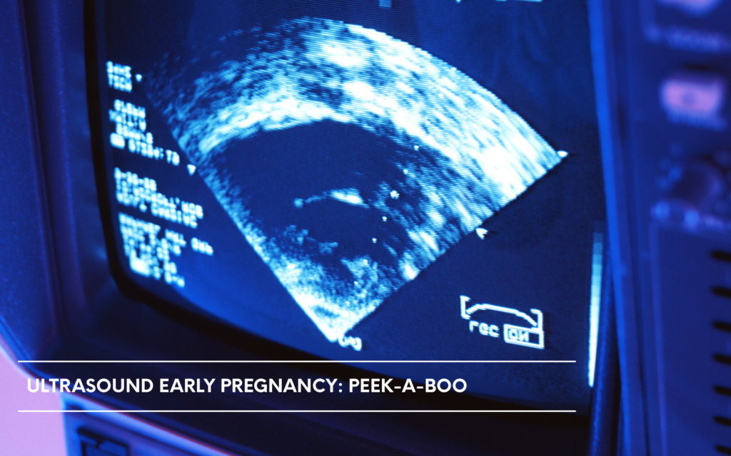
Early pregnancy ultrasounds are your first glimpse into your baby’s world. They help confirm your pregnancy, check your baby’s heartbeat, and make sure everything is developing normally. Many women feel anxious about their first scan, but knowing what to expect can help you feel more prepared and confident.
We’ll walk you through when you actually need these early scans and what happens during your appointment. You’ll also learn how to read your ultrasound results and understand what all those measurements and images really mean. Finally, we’ll address common worries that pop up during early ultrasounds so you know when something is normal and when you might need to ask more questions.
Understanding When You Need Early Pregnancy Ultrasounds
Confirming pregnancy after positive home test
That little plus sign on your pregnancy test brings excitement and questions – but home tests aren’t perfect. While modern pregnancy tests are highly accurate, they can occasionally give false positives due to certain medications, medical conditions, or even faulty tests. An early pregnancy ultrasound provides visual confirmation that you’re truly pregnant.
Your doctor will typically schedule this confirmation ultrasound around 6-8 weeks after your last menstrual period. At this stage, the gestational sac becomes visible, and you might even see a tiny flickering heartbeat. This visual confirmation gives you and your healthcare team confidence to move forward with prenatal care planning.
Some women experience early pregnancy symptoms like nausea, breast tenderness, or fatigue even before testing. When these symptoms appear alongside a positive test, an ultrasound helps connect all the dots and provides that reassuring visual proof many expectant parents need.
Determining exact gestational age
Calculating pregnancy timing based on your last menstrual period works for women with regular 28-day cycles, but many women have irregular periods or can’t remember their exact dates. Early ultrasound measurements provide the most accurate way to determine how far along you are.
During the first trimester, measuring the crown-rump length (the distance from the top of the head to the bottom of the spine) gives precise dating within 3-5 days of accuracy. This measurement is more reliable than later ultrasounds because early fetal development follows consistent patterns.
Accurate dating matters for several important reasons:
- Due date calculation: Knowing your exact due date helps plan for delivery and maternity leave
- Screening test timing: Genetic screening tests must happen within specific windows
- Monitoring development: Each week of pregnancy brings expected milestones
- Medical decision-making: Treatment options sometimes depend on exact gestational age
Healthcare providers at facilities like those in Prabhadevi and Mahim areas of Mumbai often see women who need dating ultrasounds due to irregular cycles or unclear conception timing.
Ruling out ectopic pregnancy
Ectopic pregnancy occurs when a fertilized egg implants outside the uterus, most commonly in the fallopian tubes. This serious condition affects about 1 in 50 pregnancies and requires immediate medical attention to prevent life-threatening complications.
Early pregnancy symptoms combined with certain warning signs make ectopic pregnancy a concern:
- Pelvic or abdominal pain: Sharp or cramping pain on one side
- Vaginal bleeding: Light to heavy bleeding that differs from normal periods
- Shoulder pain: Pain that radiates to the shoulder area
- Dizziness or weakness: Signs that internal bleeding might be occurring
An ultrasound can quickly identify whether the pregnancy is developing in the right location. When the gestational sac appears clearly within the uterine cavity, ectopic pregnancy is essentially ruled out. However, if the sac isn’t visible in the uterus and pregnancy hormone levels are rising, doctors investigate further for an ectopic pregnancy.
Early detection through ultrasound allows for less invasive treatment options. When caught early, medications can sometimes resolve ectopic pregnancies without surgery. Delayed diagnosis can lead to fallopian tube rupture, internal bleeding, and emergency surgery.
Assessing viability in high-risk cases
Women with certain risk factors benefit from early ultrasound monitoring to assess pregnancy viability. These risk factors include previous miscarriages, fertility treatments, advanced maternal age, or underlying health conditions.
Previous pregnancy loss creates understandable anxiety about the current pregnancy. Early ultrasounds can detect a fetal heartbeat, which significantly reduces miscarriage risk. Seeing that tiny flicker on the screen provides tremendous emotional relief for couples who’ve experienced loss before.
Fertility treatment pregnancies often involve multiple embryos or medications that increase certain risks. Women who’ve undergone IVF, IUI, or taken fertility drugs typically receive closer monitoring from the start. Early ultrasounds help detect multiple pregnancies and ensure proper development.
Bleeding during early pregnancy affects up to 25% of pregnant women and doesn’t always indicate problems. Ultrasound helps distinguish between harmless implantation bleeding and more serious issues. When the baby appears healthy with a strong heartbeat despite bleeding, most women can continue their pregnancies normally.
High-risk medical conditions like diabetes, autoimmune disorders, or blood clotting issues require careful monitoring from conception. These conditions can affect early pregnancy development, making ultrasound screening essential for appropriate care planning.
Sirona Shanti Diagnostics at Prabhadevi and Mahim communities, provide specialized early pregnancy ultrasound services for high-risk cases, ensuring expectant mothers receive proper monitoring and care.
Timing recommendations for optimal results
Your first pregnancy ultrasound typically happens between 6 to 8 weeks of pregnancy, though this can vary based on your specific situation. At 6 weeks, you’ll usually see a tiny embryo with a visible heartbeat, which many expectant parents find incredibly reassuring. If you go too early – say at 4 or 5 weeks – you might only see an empty gestational sac, which can cause unnecessary worry.
The sweet spot for that first scan is around 7-8 weeks when everything becomes much clearer. You’ll see the baby’s shape forming, hear the heartbeat loud and clear, and get accurate measurements for dating your pregnancy. Some doctors prefer waiting until 8-10 weeks, especially if you have irregular periods or aren’t sure about your last menstrual period.
If you’re experiencing bleeding, cramping, or severe morning sickness, your doctor might schedule an earlier ultrasound to check everything’s progressing normally. Emergency situations call for immediate scans regardless of timing.
Preparation steps before your appointment
Getting ready for your ultrasound is pretty straightforward, but a few simple steps can make the experience much smoother. Most early pregnancy ultrasounds require a full bladder, so you’ll need to drink about 32 ounces of water an hour before your appointment. Yes, it’s uncomfortable, but that full bladder pushes your uterus up and creates a better window for viewing your baby.
Bring your partner or support person along if you’d like. Many couples treasure this moment as their first “meeting” with their baby.
If you’re nervous (totally normal!), consider writing down questions beforehand. You might forget what you wanted to ask once you see that tiny heartbeat flickering on the screen. Quality diagnostic centers like Sirona Shanti Diagnostics in Prabhadevi and Mahim often provide comfortable environments that help ease first-time pregnancy anxieties.
Duration and comfort level of the procedure
The actual ultrasound takes about 15-30 minutes, though your entire appointment might last longer with check-in, paperwork, and consultation time. The scanning itself is completely painless – you’ll only feel the cool gel on your belly and gentle pressure from the transducer as the technician moves it around to get different views.
The most uncomfortable part is honestly that full bladder you’ve been carrying around! Once the scan starts, you’ll probably forget about the discomfort because you’ll be so focused on seeing your baby for the first time. The room is usually dimmed so you can see the monitor clearly, and most technicians will angle the screen so you can watch along.
Some women feel emotional during their first ultrasound – tears of joy, relief, or even unexpected anxiety are all completely normal reactions. The technician has seen it all and will give you space to process the moment.
You might need a transvaginal ultrasound instead of or in addition to the abdominal one, especially if you’re very early in pregnancy or if your baby is hiding behind your bladder. While this sounds intimidating, it’s actually more comfortable than the abdominal scan because you can empty your bladder first! The transvaginal probe is thin, covered with a protective sheath, and inserted only about two inches. Many women say it feels similar to a routine pap smear and provides incredibly clear, detailed images of early pregnancy.
Key Information Your Early Ultrasound Reveals
Heartbeat detection and fetal viability
The most exciting moment during your early ultrasound is hearing your baby’s heartbeat for the first time. This tiny flutter can typically be detected around 6-7 weeks of pregnancy and serves as a strong indicator of fetal viability. Your doctor will measure the heart rate, which should be between 110-160 beats per minute in early pregnancy. A strong, regular heartbeat significantly reduces the risk of miscarriage and confirms that your pregnancy is developing normally.
Number of babies you’re carrying
Your early ultrasound quickly reveals whether you’re expecting one baby or multiples. The sonographer will count the number of gestational sacs and embryos present. If you’re carrying twins or more, they’ll also determine if they’re identical or fraternal by examining the placental and membrane structures. This information is crucial for planning your prenatal care, as multiple pregnancies require more frequent monitoring and specialized medical attention.
Placental position and health
The ultrasound provides valuable insights into your placenta’s location and development. Your healthcare provider will check for proper implantation and ensure the placenta is forming correctly. They’ll look for any signs of placenta previa (when the placenta covers the cervix) or other positioning issues that might affect your pregnancy. Early detection of placental concerns allows for appropriate monitoring and management throughout your pregnancy journey.
Estimated due date accuracy
Your early ultrasound offers the most accurate way to determine your due date. By measuring the crown-rump length of your baby, doctors can calculate gestational age within just a few days of accuracy. This measurement is more reliable than using your last menstrual period, especially if you have irregular cycles. An accurate due date helps guide your entire prenatal care schedule and ensures proper timing for important tests and appointments.
Early detection of potential complications
Early pregnancy ultrasounds can identify various conditions that require monitoring or intervention. These include ectopic pregnancies (where the embryo implants outside the uterus), molar pregnancies, or chromosomal abnormalities. Your ultrasound technician will also check for proper organ development and look for any structural abnormalities. While detecting these issues early can be concerning, it allows your medical team at facilities like Sirona Shanti Diagnostics in Prabhadevi, Mumbai to provide timely care and support, ensuring the best possible outcomes for both you and your baby.
Types of Early Pregnancy Ultrasound Procedures
Transvaginal Ultrasound Benefits and Process
Transvaginal ultrasounds are the gold standard for early pregnancy imaging, particularly during the first trimester. This procedure involves inserting a specialized ultrasound probe into the vagina, allowing doctors to get incredibly clear images of the developing embryo or fetus. The probe sits closer to your uterus than an abdominal ultrasound would, which means much sharper, more detailed pictures.
The process is straightforward and typically takes 15-20 minutes. You’ll lie on an examination table with your knees bent, similar to a gynecological exam. The ultrasound technician covers the probe with a protective cover and lubricating gel before gentle insertion. While some women feel mild discomfort, the procedure isn’t painful for most people.
The biggest advantage of transvaginal ultrasounds is their accuracy in early pregnancy. They can detect pregnancy as early as 4-5 weeks, when abdominal ultrasounds might not show anything yet. You’ll get precise measurements of the gestational sac, clear views of the baby’s heartbeat, and accurate dating of your pregnancy. This type of scan also helps identify potential complications like ectopic pregnancies or miscarriages much earlier than other methods.
Abdominal Ultrasound Timing and Limitations
Abdominal ultrasounds work best after 8-10 weeks of pregnancy when the uterus has grown large enough to rise above the pelvic bone. During this procedure, you’ll lie on your back while the technician applies gel to your belly and moves a transducer across your skin. The sound waves bounce back to create images on a monitor.
The main limitation of abdominal ultrasounds early in pregnancy is image quality. Before 8 weeks, the developing baby is still very small and sits deep in the pelvis, making it difficult to see clearly through the abdominal wall. Your bladder, intestinal gas, and body weight can also affect image clarity.
You’ll need a full bladder for most early abdominal ultrasounds, which helps push the uterus up and provides a clearer window for imaging. While this can be uncomfortable, it significantly improves the quality of the pictures. Many women prefer abdominal ultrasounds because they’re less invasive than transvaginal procedures.
| Ultrasound Type | Best Timing | Image Quality | Preparation Needed |
|---|---|---|---|
| Transvaginal | 4-12 weeks | Excellent | Empty bladder |
| Abdominal | 8+ weeks | Good to excellent | Full bladder |
3D and 4D Ultrasound Options
Three-dimensional (3D) and four-dimensional (4D) ultrasounds offer exciting glimpses of your baby, though they’re not typically necessary for routine early pregnancy care. 3D ultrasounds create detailed, lifelike images that look like photographs, while 4D ultrasounds show real-time movement, letting you see your baby yawning, moving their hands, or even sucking their thumb.
These advanced imaging techniques work best between 24-34 weeks of pregnancy when there’s enough amniotic fluid around the baby and facial features are well-developed. Before 20 weeks, the images might not be as clear or recognizable as you’d hope.
While 3D and 4D ultrasounds are incredibly popular for bonding and keepsake purposes, they don’t provide additional medical information beyond what standard 2D ultrasounds offer. Most doctors use them as supplementary tools rather than primary diagnostic methods. The sessions typically last 30-45 minutes and often result in beautiful photos and videos that families treasure.
Some diagnostic centers, including specialized facilities in areas like Prabhadevi and Mahim in Mumbai, offer these advanced ultrasound services as part of comprehensive pregnancy care packages. When choosing a provider, look for certified technicians and modern equipment to ensure the best possible experience and image quality.
Normal Measurements and Developmental Milestones
Early pregnancy ultrasounds provide specific measurements that help track your baby’s healthy development. Between 6-8 weeks, you’ll see the gestational sac measuring approximately 10-40mm in diameter. The yolk sac should appear round and measure 3-6mm, serving as your baby’s first source of nutrition. By week 6, the fetal pole becomes visible as a small curved structure measuring 2-4mm.
Your healthcare provider will compare these measurements against standardized growth charts. At 7 weeks, you should detect a fetal heartbeat ranging from 100-160 beats per minute. The embryo typically measures 4-10mm at this stage. Each week brings new milestones – limb buds appear around week 8, and basic organ formation begins.
These measurements help confirm your pregnancy is progressing normally and can detect potential complications early. Your doctor will plot these numbers on growth curves to ensure your baby develops at the expected pace.
Understanding Gestational Sac Appearance
The gestational sac is your pregnancy’s first visible sign on ultrasound, typically appearing around 4.5-5 weeks after your last menstrual period. This fluid-filled structure looks like a small, dark circle surrounded by a bright white rim called the decidual reaction.
A healthy gestational sac should appear round or oval-shaped with smooth, well-defined borders. The sac grows approximately 1mm per day during early pregnancy. By week 5, it measures about 10-15mm in diameter. The shape and growth rate provide valuable information about pregnancy viability.
Irregular sac shapes, poorly defined borders, or slower-than-expected growth may indicate potential concerns. Your ultrasound technician will examine the sac’s position within the uterus to confirm proper implantation. The sac should be centrally located in the upper portion of your uterus, embedded in the thickened endometrial lining.
Crown-Rump Length Significance
Crown-rump length (CRL) measures your baby from the top of the head to the bottom of the rump, providing the most accurate dating method during early pregnancy. This measurement becomes possible once the fetal pole reaches about 5mm, typically around 6-7 weeks gestation.
CRL measurements are incredibly precise for pregnancy dating, with accuracy within 3-5 days when measured between 7-13 weeks. Your baby grows approximately 1mm per day during this period. At 8 weeks, the CRL measures roughly 16mm, growing to about 30mm by week 10.
Healthcare providers use CRL to establish your estimated due date, especially when your last menstrual period date is uncertain. This measurement also helps identify potential growth concerns or chromosomal abnormalities. Significant deviations from expected CRL measurements may prompt additional testing or monitoring.
The CRL provides more reliable dating than using your last menstrual period, particularly if you have irregular cycles or aren’t sure about conception dates.
When Follow-Up Scans Are Recommended
Several situations warrant additional ultrasound appointments beyond your routine first scan. If your initial ultrasound shows an empty gestational sac or measurements smaller than expected, your doctor will schedule a follow-up scan in 1-2 weeks to monitor development.
Bleeding during early pregnancy requires repeat scanning to check fetal viability and rule out miscarriage or ectopic pregnancy. Previous pregnancy complications, such as miscarriage or ectopic pregnancy, often necessitate more frequent monitoring through additional scans.
When initial measurements don’t match your expected due date by more than a week, follow-up scans help clarify accurate dating. Multiple pregnancies detected during your first scan require ongoing monitoring to track each baby’s development.
Facilities like Sirona Shanti Diagnostics in Prabhadevi, Mumbai, provide comprehensive follow-up care when additional monitoring is needed. They offer flexible scheduling to accommodate urgent concerns or routine follow-up appointments.
Certain medical conditions, including diabetes, thyroid disorders, or fertility treatment pregnancies, typically require more frequent ultrasound monitoring. Your healthcare provider will create a personalized scanning schedule based on your specific circumstances and risk factors.
Common Concerns and What They Mean
Empty Gestational Sac Explanations
An empty gestational sac, also known as a blighted ovum or anembryonic pregnancy, occurs when a fertilized egg implants in the uterus but fails to develop into an embryo. This situation affects roughly 15-20% of all pregnancies and can be emotionally challenging for expecting parents.
During your early ultrasound, the technician looks for specific structures within the gestational sac. When the sac appears empty without visible fetal pole or yolk sac, several scenarios might explain this finding:
- Too early for detection: If you’re very early in pregnancy (before 6 weeks), the embryo might simply be too small to visualize yet
- Dating discrepancy: Your conception date might be different from what you calculated, making the pregnancy appear less advanced
- True blighted ovum: The pregnancy stopped developing after implantation but before embryo formation
Your healthcare provider will likely recommend follow-up scans 1-2 weeks later to confirm the diagnosis. Rising or declining hCG levels also provide valuable information about pregnancy viability.
Slow Fetal Heart Rate Implications
Fetal bradycardia, or slow heart rate, can raise concerns during early pregnancy ultrasounds. Normal fetal heart rates range from 120-160 beats per minute after 8 weeks, but early detection around 6-7 weeks often shows lower rates of 90-120 bpm.
Several factors influence early fetal heart rate:
- Gestational age accuracy: Earlier pregnancies naturally have slower heart rates
- Measurement timing: Heart rate can fluctuate during the scan
- Technical factors: Equipment sensitivity and fetal position affect readings
A consistently slow heart rate below 90 bpm after 8 weeks may indicate:
- Chromosomal abnormalities
- Maternal health conditions affecting blood flow
- Placental insufficiency
- Impending pregnancy loss
Your doctor will monitor heart rate trends over multiple visits rather than making decisions based on a single measurement. Serial scans help distinguish between normal variation and concerning patterns.
Bleeding During Early Pregnancy Assessment
Spotting or bleeding affects nearly 25% of pregnant women during their first trimester, creating anxiety about pregnancy viability. Ultrasound evaluation helps determine the source and significance of early pregnancy bleeding.
Types of bleeding your ultrasound can identify:
| Bleeding Type | Ultrasound Findings | Implications |
|---|---|---|
| Implantation | Normal sac placement | Usually benign |
| Subchorionic hematoma | Blood collection near placenta | Variable outcomes |
| Threatened abortion | Closed cervix, viable pregnancy | Monitoring required |
| Inevitable abortion | Open cervix | Pregnancy loss likely |
Assessment factors include:
- Amount and color of bleeding
- Cervical opening status
- Fetal heart activity
- Gestational sac appearance
- Presence of blood clots or hematomas
Light spotting with normal ultrasound findings often resolves without intervention. However, heavy bleeding accompanied by cramping and abnormal scan results requires immediate medical attention.
Healthcare providers at facilities like those in Prabhadevi and Mahim areas of Mumbai use advanced ultrasound technology to accurately assess bleeding causes and provide appropriate guidance. Early intervention and proper monitoring significantly improve outcomes for pregnancies complicated by bleeding episodes.
Your medical team will create a customized monitoring plan based on your specific ultrasound findings, symptoms, and risk factors.
Getting an early pregnancy ultrasound can feel overwhelming, but knowing what to expect makes the experience much smoother. These scans give you and your doctor important information about your baby’s development, help confirm your due date, and can catch any potential issues early on. The different types of procedures each serve specific purposes, and your healthcare provider will recommend the right approach based on your individual situation.
Remember that every pregnancy is unique, and what you see on your ultrasound might look different from what friends or family experienced. Don’t hesitate to ask your technician or doctor questions during your appointment – they’re there to help you understand what you’re seeing and address any worries you might have. Trust the process, stay informed about what each scan reveals, and enjoy these first glimpses of your growing baby.
Common Keywords:
Best NABL accredited pathology lab in Mumbai, Best pathology lab in Mahim Dadar Prabhadevi Worli, Best radiology centre center in mumbai, Best sonography center in mumbai, Best Radiologist / Sonologist in Mahim Worli Dadar Prabhadevi, Best Cardiologist in Mumbai, best cardiology centre, centre in Mahim Dadar Prabhadevi, ecg at home, xray at home, blood collection at home, blood collection in mumbai, bloold collection south mumbai, verified best diagnostics centers
Disclaimer: The information on this site is just meant to be used for learning and general knowledge. It does not take the place of professional medical advice, diagnosis, or care. If you need personalised medical advice, go to a diagnostic centre or talk to a qualified healthcare practitioner.
Dr. Kunaal Jain
Consultant Radiologist