

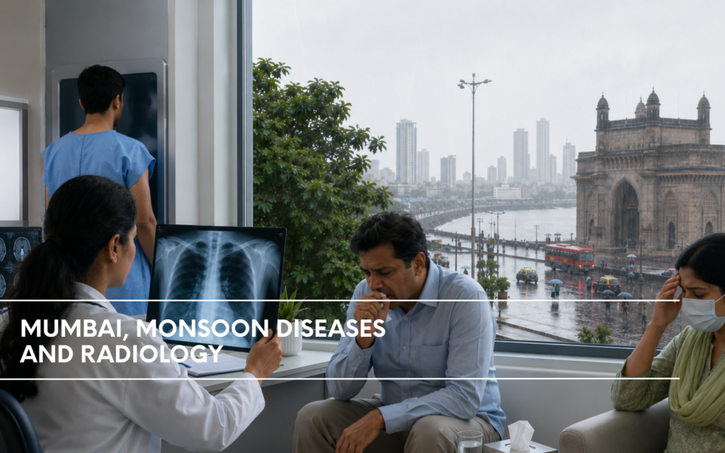
Mumbai, Monsoon Diseases and Radiology
By Dr. Riya Kothari Jain, Radiologist | Sirona Shanti Diagnostics, Mumbai
Every year, as the southwest monsoon rolls in over the Arabian Sea and drenches Mumbai in its first heavy downpour, our reporting room at Sirona Shanti Diagnostics quietly braces for what we in radiology know as “the season.” The city erupts in joy, the heat breaks, the streets fill with the smell of wet earth but within days, our radiology work list begins to tell a different story. Chest X-rays of patients with rattling coughs. Ultrasound abdomen scans of children with tender, enlarged livers. Soft tissue swellings. Flooded sinuses. The monsoon in Mumbai is not just a meteorological event. For those of us reading images in a diagnostic centre, it is a clinical season unto itself.
Over the years, I have come to appreciate how powerfully imaging shapes the management of monsoon-related illness. Clinicians suspect; we confirm, refine, or redirect. This blog is my attempt to walk through the major diseases that flood our worklist every June through September, and explain what we see and why it matters on X-ray and ultrasound.
Why Mumbai Is a Perfect Storm for Monsoon Illness
Before we dive into individual diseases, it helps to understand why Mumbai is particularly vulnerable. The city’s geography low-lying, densely populated, crisscrossed by open drains and tidal creeks means that heavy rainfall rapidly contaminates drinking water sources. Waterlogging turns streets into wading pools. Humidity soars to 85–95%. Stagnant water breeds mosquitoes within 48 hours. Crowded local trains and chawls accelerate respiratory transmission. Malnutrition and suboptimal sanitation in several pockets of the city lower immune defences.
The result: every monsoon season brings predictable surges in leptospirosis, typhoid, malaria, dengue, hepatitis A and E, gastroenteritis, and respiratory infections. As a radiologist, I see the downstream consequences of all of these.
1. Leptospirosis Mumbai’s Most Feared Monsoon Killer
The Clinical Background
Leptospirosis is caused by Leptospira interrogans, a spirochete shed in the urine of rats, dogs, and cattle all present in abundance across Mumbai’s flooded streets. Wading through knee-deep floodwater, which is virtually unavoidable during heavy rains, allows the organism to enter through skin abrasions or intact mucous membranes. Mumbai has recorded some of the highest case fatalities from leptospirosis in India, particularly during severe flood years.
The disease ranges from a mild flu-like illness (Weil’s syndrome precursor) to the full triad of jaundice, acute kidney injury, and pulmonary haemorrhage, the dreaded Weil’s disease.
What We See on Chest X-Ray
Pulmonary involvement in leptospirosis is one of the most dramatic things I encounter on a chest radiograph during the monsoon season. The mechanism is direct endothelial damage by the spirochete, causing diffuse alveolar haemorrhage (DAH), not pneumonia in the traditional sense.
On the chest X-ray, we see:
-
Bilateral alveolar opacities are typically basal-predominant in early disease, but rapidly become diffuse as DAH worsens. These opacities are fluffy, confluent, and have no lobar or segmental boundaries, which distinguishes them from consolidative pneumonia.
-
Air bronchograms visible within the opacified areas, confirming that the airways themselves are patent while the alveoli fill with blood or fluid.
-
Perihilar “bat-wing” pattern in severe cases, opacities radiate outward from the hila, resembling pulmonary oedema. This creates a diagnostic challenge: is this cardiogenic pulmonary oedema, leptospiral lung, or ARDS? Clinical context, serology, and the absence of cardiomegaly or Kerley B lines on the radiograph help steer the diagnosis.
-
Pleural effusions and small bilateral effusions are not uncommon.
- In cases where the patient deteriorates to ARDS, the radiograph becomes increasingly “whited out,” with dense bilateral opacification. At this stage, we are monitoring rather than diagnosing looking for complications like pneumothorax from barotrauma in ventilated patients.
What We See on Ultrasound
While chest X-ray handles the pulmonary picture, ultrasound is invaluable for assessing the abdominal organs, particularly in Weill’s disease.
-
Hepatomegaly the liver is enlarged and may be heterogeneously echogenic due to diffuse hepatocellular injury. Jaundice in leptospirosis is hepatocellular in origin, and the liver parenchyma often looks brighter than normal due to inflammation.
-
Acute kidney injury kidneys appear enlarged and edematous, with increased cortical echogenicity and reduced corticomedullary differentiation. These findings reflect tubular necrosis, the primary renal injury in leptospirosis.
-
Gallbladder wall thickening a subtle but consistent finding. The gallbladder wall becomes edematous and layered, sometimes with pericholecystic fluid. This is not cholecystitis; it reflects diffuse capillary leak and hypoalbuminemia.
-
Ascites free fluid in the abdomen, particularly in the hepatorenal space (Morison’s pouch) and the pelvis, indicates third-spacing from hypoalbuminemia and capillary injury.
-
Splenomegaly mild to moderate splenic enlargement is common and reflects reticuloendothelial activation.
- Point-of-care ultrasound in the emergency setting has become invaluable for rapid triaging of leptospirosis patients. Finding pleural effusions, ascites, and kidney changes together in a febrile patient who waded through floodwater is a strong prompt to initiate empiric treatment while awaiting confirmatory serology.
2. Typhoid Fever The Waterborne Silent Invader
The Clinical Background
Salmonella typhi thrives in Mumbai’s monsoon because flooding contaminates water supply lines and open wells. Typhoid remains endemic, but case numbers rise sharply from July to September. The classic step-ladder fever, relative bradycardia, and “rose spots” are not always present in clinical practice; more commonly, patients present with prolonged fever, headache, and abdominal discomfort.
What We See on Chest X-Ray
Pulmonary involvement is less prominent in typhoid than in leptospirosis, but it does occur:
-
Lobar pneumonia pattern Salmonella can cause pneumonia directly, though this is uncommon. More often, we see a secondary bacterial pneumonia complicating typhoid.
-
Relative absence of findings importantly, the chest X-ray is often unremarkable in uncomplicated typhoid. Its value lies in excluding alternative diagnoses: malaria, leptospirosis, or TB, which may mimic typhoid clinically.
-
Chest X-ray for complications of intestinal perforation is the most feared complication of typhoid. While CT is the gold standard for perforation, an erect chest X-ray may show free air under the diaphragm (pneumoperitoneum). In our setting, when a typhoid patient acutely deteriorates with abdominal rigidity, an erect CXR is the first step before escalating to CT.
What We See on Ultrasound
Ultrasound is central to typhoid management both for diagnosis and complication surveillance.
-
Hepatosplenomegaly is almost universal in typhoid fever. The liver and spleen are diffusely enlarged, with normal to mildly altered echotexture. Measuring the organ dimensions serially on follow-up helps gauge response to treatment.
-
Mesenteric lymphadenopathy is one of the most consistent ultrasound findings in typhoid. We see multiple enlarged lymph nodes in the right iliac fossa and around the terminal ileum, measuring 1–2 cm, with a maintained echogenic hilum. This distribution reflects the Peyer’s patches of the terminal ileum, which are the primary site of S. typhi infection.
-
Thickened terminal ileum wall, wall thickening of the terminal ileum on ultrasound (>3 mm), along with surrounding mesenteric fat stranding echogenicity, confirms bowel wall inflammation. This is a subtle finding but consistent in my experience.
-
Gallbladder changes the gallbladder as a reservoir for S. typhi in chronic carriers. Sludge or wall thickening may be seen. In the acute carrier state, we look for cholecystitis-like changes.
-
Perforation detection when intestinal perforation occurs, ultrasound may show free peritoneal fluid, loculated collections, or air artefact in the peritoneum. However, free air is better detected on an erect CXR or CT, and perforation with peritonitis demands immediate surgical referral rather than extended imaging.
3. Malaria : The Mosquito’s Gift to the Monsoon City
The Clinical Background
Mumbai’s stagnant water pools following rain become ideal breeding grounds for Anopheles mosquitoes. Both Plasmodium vivax (the predominant species in Mumbai) and Plasmodium falciparum (the dangerous species) are endemic. Falciparum malaria, though less common, carries the risk of cerebral malaria, severe anaemia, and multiorgan dysfunction.
Radiology does not diagnose malaria, which is the blood smear’s domain. But we play a crucial role in assessing complications.
What We See on Chest X-Ray
-
Pulmonary edema / ARDS pattern severe falciparum malaria can cause non-cardiogenic pulmonary edema (malarial lung), presenting on CXR as bilateral alveolar opacities with no cardiomegaly, mimicking ARDS. Distinguishing this from leptospiral lung or viral pneumonia requires clinical and serological correlation.
-
Pleural effusions small bilateral pleural effusions from hypoalbuminemia and capillary leak are common in severe malaria.
-
Cardiomegaly malarial myocarditis, while rare, can present with an enlarged cardiac silhouette and pulmonary vascular redistribution on CXR.
What We See on Ultrasound
-
Splenomegaly is the most consistent abdominal finding. In P. vivax malaria, the spleen can become massively enlarged, sometimes reaching the iliac fossa with repeated infections (tropical splenomegaly syndrome). On ultrasound, the spleen shows increased echogenicity due to hemosiderin deposition. Measuring splenic length (>13 cm is enlarged) is part of every malarial follow-up.
-
Splenic complications spontaneous splenic rupture is rare but catastrophic. In a febrile malarial patient who suddenly develops left hypochondriac pain and hypotension, an emergency ultrasound is life-saving. We look for perisplenic free fluid, subcapsular hematoma (a hypoechoic crescent around the spleen), or frank splenic laceration.
-
Hepatomegaly the liver is enlarged with heterogeneous echotexture, and Kupffer cell activation gives it a slightly brighter appearance. Malarial pigment (hemozoin) deposits can occasionally be seen as coarse echogenicity.
-
Ascites and gallbladder thickening as in leptospirosis, third-spacing from hypoalbuminemia causes peritoneal fluid and edematous gallbladder walls.
4. Dengue Fever The Bone-Crusher and the Bleeder
The Clinical Background
Aedes aegypti, the dengue mosquito, breeds in clean stagnant water flower pots, cooler trays, and water storage containers and thus thrives in urban Mumbai even without flooding. Dengue surges every monsoon, often as the season’s dominant vector-borne disease in terms of sheer case numbers. The concern is not so much the classical dengue fever but dengue hemorrhagic fever (DHF) and dengue shock syndrome (DSS), where capillary leak and thrombocytopenia become life-threatening.
What We See on Chest X-Ray
-
Pleural effusions are the hallmark of DHF on imaging. Capillary leak causes right-sided (or bilateral) pleural effusions, which we detect as blunting of the costophrenic angle on PA CXR. Serial CXR helps track the critical phase: effusions typically worsen around days 3–6 of illness (the “leaking phase”) and resolve in the recovery phase.
-
Right-sided predominance dengue pleural effusions have a known right-sided predominance, a finding that, while not pathognomonic, is clinically useful. When I see a right-dominant effusion in a febrile patient during monsoon, dengue climbs high in my differential.
-
Pulmonary infiltrates in severe dengue with ARDS or secondary bacterial pneumonia, bilateral alveolar opacities appear. These carry a poor prognosis.
-
Cardiomegaly and pulmonary oedema dengue myocarditis, though uncommon, can cause cardiac enlargement and pulmonary vascular congestion. Recognising this on CXR is important for fluid management, as aggressive IV fluids are dangerous in dengue with cardiac involvement.
What We See on Ultrasound
Ultrasound is the workhorse investigation in dengue more so than in almost any other monsoon disease.
-
Pleural effusions are far more sensitive than CXR for small effusions. We can detect as little as 50–100 mL of pleural fluid on ultrasound, versus the 300 mL needed for costophrenic angle blunting on CXR. Right-sided effusions in dengue appear as anechoic crescents above the right hemidiaphragm.
-
Ascites free fluid in the peritoneum is a critical sign of plasma leakage in DHF. We find it first in the hepatorenal space and the pelvis. The presence of ascites with pleural effusion and a platelet count < 100,000/µL is essentially diagnostic of DHF in the right clinical context.
-
Gallbladder wall thickening is one of the earliest and most consistent ultrasound findings in dengue. The gallbladder wall becomes thickened (>3 mm) and edematous, with a characteristic layered or “double-wall” appearance, reflecting capillary leak. This finding appears early in the illness sometimes before effusions develop and serves as an early warning sign.
-
Hepatosplenomegaly both organs enlarge, though this is less dramatic than in malaria or leptospirosis. Hepatomegaly with periportal hyperechogenicity (bright halo around portal tracts) reflects periportal edema and hepatocellular injury.
-
Hemorrhagic complications in severe DHF with thrombocytopenia, ultrasound may detect haemorrhage: retroperitoneal hematoma appears as a heterogeneous collection posterior to the kidneys; hepatic hematoma as a heterogeneous intralesional collection; or pelvic hematoma. These are rare but serious findings that change surgical management.
-
Fluid monitoring during critical phases we perform serial ultrasounds (sometimes twice daily) in hospitalised DHF patients to guide fluid management. Increasing ascites or pleural effusion signals ongoing leakage; their resolution marks recovery. This real-time feedback is something no blood test can provide.
5. Hepatitis A and E The Jaundice Epidemics
The Clinical Background
Hepatitis A (HAV) and Hepatitis E (HEV) are both transmitted by the faecal-oral route through contaminated water, and both surge every monsoon in Mumbai when waterlogging contaminates the water supply. Children and young adults are predominantly affected by HAV; HEV is particularly dangerous in pregnant women, where it carries a high maternal mortality. Outbreaks often cluster in slums or areas with poor sanitation.
What We See on Ultrasound
Chest X-ray has no established role in acute viral hepatitis. Ultrasound, however, is the primary modality.
-
Hepatomegaly with altered echogenicity the liver is enlarged in the acute phase. Classically, the liver parenchyma appears hypoechoic (darker than normal) due to diffuse cellular oedema and inflammation, in contrast to the hyperechoic appearance of fatty liver or cirrhosis. This “starry sky” appearance of bright portal tracts against a dark parenchymal background is a well-recognised ultrasound sign of acute hepatitis, though not specific to HAV or HEV.
-
Gallbladder wall thickening present in acute hepatitis as well, secondary to hypoalbuminemia and periportal inflammation. The wall appears edematous and thickened (>3 mm) without tenderness or pericholecystic fluid (which would suggest cholecystitis).
-
Porta hepatis lymphadenopathy mildly enlarged lymph nodes around the porta hepatis are common in acute viral hepatitis, reflecting reactive hyperplasia.
-
Splenomegaly the spleen enlarges modestly, reflecting portal hypertension from the acute hepatic inflammation. Severe or persistent splenomegaly beyond the acute phase raises concern for evolving portal hypertension.
-
Doppler evaluation in severe hepatitis approaching acute liver failure, portal vein Doppler is important. We look for portal vein thrombosis (absent flow or echogenic thrombus), hepatic vein waveform changes, or reversed portal flow, all indicators of severe parenchymal dysfunction.
-
Differentiating from obstructive jaundice, perhaps the most critical role of ultrasound in a jaundiced patient is excluding biliary obstruction. In acute viral hepatitis, the biliary system is typically normal: the CBD is not dilated, there are no stones or masses. This simple finding redirects the entire workup away from surgery and toward medical management.
6. Acute Gastroenteritis The Most Common, Often Underestimated
The Clinical Background
Acute gastroenteritis from contaminated food and water peaks every monsoon. Pathogens include Vibrio cholerae, E. coli, Shigella, Cryptosporidium, and Giardia. While most cases are self-limiting, severe dehydration particularly in children, the elderly, and immunocompromised patients can be fatal. Cholera outbreaks, though less frequent than historically, still occur in Mumbai’s peri-urban areas.
The Role of Imaging
For most acute gastroenteritis, imaging is not required, clinical assessment and stool culture suffice. However, radiology becomes crucial in specific scenarios:
Chest X-ray for cholera complications severe cholera can cause aspiration pneumonia (from recumbent vomiting) or secondary pulmonary oedema from fluid resuscitation. CXR guides fluid management in complicated cases.
Ultrasound for complications and differentials:
- Intussusception in children with viral gastroenteritis (especially adenovirus and rotavirus), intussusception is a feared complication. Ultrasound is the investigation of choice, showing the classic “target sign” or “pseudo-kidney sign” in transverse and longitudinal views, respectively. Early diagnosis can allow pneumatic or hydrostatic reduction, avoiding surgery.
- Appendicitis mimicking gastroenteritis one of the most common and consequential pitfalls in monsoon gastroenteritis is missing an acute appendicitis. We see this regularly: a patient with fever, vomiting, and “loose stools” gets labelled as gastroenteritis; ultrasound is requested for abdominal pain that doesn’t resolve. We look for a non-compressible, aperistaltic tubular structure >6 mm in the right iliac fossa, with periappendiceal fat hyperechogenicity and free fluid. Catching this early makes the difference between a laparoscopic appendicectomy and a perforated appendicitis with peritonitis.
- Paralytic ileus / small bowel obstruction in severe or prolonged gastroenteritis, the bowel may become adynamic. Ultrasound shows dilated, fluid-filled loops of bowel with absent or decreased peristalsis. Distinguishing this from mechanical obstruction (fixed, non-peristaltic loop with abrupt transition point) sometimes requires a supine abdominal X-ray to identify air-fluid levels.
- Mesenteric lymphadenitis clusters of enlarged mesenteric lymph nodes in the right iliac fossa, sometimes indistinguishable from early typhoid lymphadenopathy, are seen in non-specific mesenteric adenitis associated with viral gastroenteritis. This is a reassuring finding that argues against a surgical cause of abdominal pain.
7. Respiratory Infections The Airway’s Monsoon Burden
The Clinical Background
Humid monsoon air, crowded transport, and increased indoor time accelerate the transmission of respiratory pathogens influenza viruses, Streptococcus pneumoniae, Haemophilus influenzae, and Mycobacterium tuberculosis (whose reactivation risk increases with malnutrition during the monsoon). Community-acquired pneumonia (CAP), acute exacerbations of asthma and COPD, and viral upper respiratory infections dominate the outpatient respiratory load.
The Role of Chest X-Ray
Chest radiography remains the first-line and most critical imaging modality for respiratory infections.
-
Lobar/segmental consolidation bacterial pneumonia, particularly pneumococcal pneumonia, presents as homogeneous opacification of a lobe or segment, with air bronchograms and no volume loss. The lower lobes are most commonly affected. On PA and lateral views, we identify the affected lobe and guide antibiotic choice.
-
Bronchopneumonia pattern patchy, bilateral, peribronchial opacities indicate bronchopneumonia, typically from Staphylococcus, Haemophilus, or Klebsiella. These lack the lobar homogeneity of pneumococcal CAP.
-
Interstitial pattern fine bilateral reticular or ground-glass opacities suggest atypical pneumonia (Mycoplasma, Chlamydophila, viral pneumonia). The chest X-ray often looks worse than the patient, a classic feature of atypical pneumonia.
-
Parapneumonic effusion and empyema blunting of the costophrenic angle indicates a pleural effusion. Ultrasound is superior to CXR for characterising this: is it simple (anechoic, free-flowing) or complex (septated, echogenic)? A complex parapneumonic effusion or empyema requires drainage; simple effusions often resolve with antibiotics alone. I find that ultrasound-guided thoracocentesis is both safer and more successful than blind drainage, and our team at Sirona Shanti performs this routinely.
-
Tuberculosis Mumbai is a high-TB-burden city. Every monsoon, we encounter patients with coughs labeled as “seasonal infection” who harbor active TB. I maintain a high index of suspicion for TB whenever I see: upper lobe heterogeneous opacities, cavitation, unilateral hilar or paratracheal lymphadenopathy, or unilateral pleural effusion. In a febrile patient, TB and community-acquired pneumonia can be indistinguishable on CXR at presentation and that diagnostic uncertainty is precisely where sputum smear and GeneXpert become critical.
-
Ultrasound for respiratory infections lung ultrasound (LUS) is increasingly used at the bedside to assess pneumonia, effusions, and pulmonary oedema. We detect B-lines (vertical reverberation artefacts from subpleural alveolar fluid), consolidation (tissue-like echogenicity with air bronchograms), and effusions. In children especially, where radiation concerns make repeated CXR undesirable, LUS is a valuable tool.
8. Leptospiral Uveitis The Delayed Eye Complication
This deserves a brief mention because it is specific to leptospirosis and often missed: weeks to months after the acute infection, patients can develop uveitis from immune-mediated ocular inflammation. While this is an ophthalmology domain, patients sometimes present with ocular symptoms during the period when radiologists are still seeing the residual chest and abdominal findings. Being aware of this late complication reminds us that leptospirosis is not just an acute monsoon event; it has a long tail.
9. Soft Tissue Infections The Wading-Water Wounds
The Clinical Background
Wading through contaminated floodwater exposes cuts, abrasions, and even intact skin to polymicrobial contamination. We see an uptick in cellulitis, subcutaneous abscesses, infected diabetic foot ulcers, and in severe cases necrotizing fasciitis (NF) during the monsoon.
The Role of Ultrasound and X-Ray
Ultrasound for soft tissue infections this is an area where ultrasound directly changes surgical decision-making. In a swollen, erythematous limb:
- Simple cellulitis: diffuse subcutaneous oedema (cobblestone appearance hyperechoic fat lobules separated by fluid-filled anechoic lines). No drainable collection. Medical management.
- Abscess: a well-defined hypoechoic or anechoic collection, often with internal echoes (pus), posterior acoustic enhancement, and surrounding hyperemia on Doppler. Ultrasound-guided aspiration or drainage is the treatment. I have seen numerous cases where clinical palpation missed a deep abscess that ultrasound clearly identified.
- Necrotising fasciitis: the most dangerous diagnosis to miss. On ultrasound, we look for fascial thickening, fluid tracking along fascial planes, and the pathognomonic finding gas within the soft tissues appearing as bright hyperechoic foci with “dirty shadowing” and reverberation artefact. This gas is produced by anaerobic bacteria. Finding gas in the fascial planes is a surgical emergency; the patient needs immediate debridement, not more imaging.
X-ray for soft tissue gas a plain radiograph of the involved limb can show gas in the soft tissues as linear radiolucencies tracking along fascial planes. This is a quick, low-cost red flag for necrotising infection.
A Word on Our Workflow at Sirona Shanti Diagnostics
Working at Sirona Shanti Diagnostics gives us the advantage of combining digital radiography with high-resolution ultrasound in an accessible, patient-centred environment. During the monsoon months, our team often sees patients who have been triaged from nearby clinics or hospitals, and turnaround time on imaging reports becomes clinically critical.
A few principles guide our monsoon radiology workflow:
Integrated reporting: When we receive a chest X-ray for “monsoon fever,” we do not read it in isolation. We correlate the request form’s clinical details flood exposure, duration of fever, bleeding tendency, jaundice to calibrate our differential. A bilateral basal opacity means something different in a flood worker with jaundice (leptospirosis lung) versus a child in a housing society (viral pneumonia).
Opportunistic ultrasound: When a patient comes in for an abdominal ultrasound, we routinely assess the pleural spaces, gallbladder wall, and free fluid findings that shift the diagnosis even when not explicitly requested. A three-minute sweep of the thorax for pleural effusions has changed clinical management for many dengue and leptospirosis patients in my experience.
Communication with the treating physician: For critical findings pneumoperitoneum on CXR, gas in soft tissues on X-ray, perisplenic hematoma on ultrasound we call the referring doctor immediately. Radiology reports are only useful if they reach the right hands at the right time.
Avoiding over-reliance on imaging in clear clinical cases: Not every monsoon fever needs a chest X-ray. Not every abdominal pain needs an ultrasound. We counsel referring clinicians on appropriate imaging indications to avoid unnecessary radiation and cost, and to keep our reporting queue focused on cases where imaging genuinely adds value.
The Broader Picture: Radiology as the Monsoon’s Quiet Chronicler
There is something quietly profound about working as a radiologist in Mumbai during the monsoon. Every image that passes through our workstation tells the story of the city’s struggle with its own geography, its inadequate drainage, its dense population, its resilient but strained public health infrastructure. The X-ray of a flood worker’s lungs is white with haemorrhage. The ultrasound of a child’s liver shows swelling with hepatitis. The abdominal scan of a pregnant woman with fulminant HEV.
We do not treat these patients. But we illuminate their illness. We answer the clinical questions that cannot be answered at the bedside alone. We distinguish the surgical from the medical emergency, the viral from the bacterial, the benign from the ominous.
At Sirona Shanti Diagnostics, our commitment during the monsoon as throughout the year is to be that reliable, rapid, and thoughtful imaging partner for Mumbai’s clinicians and their patients. Because in medicine, as in the monsoon itself, timing is everything.
The author practices radiology at Sirona Shanti Diagnostics, Mumbai. All clinical impressions described reflect general radiological observations and should be interpreted in the context of the patient’s full clinical picture. Imaging findings alone do not constitute a diagnosis.
For appointments and emergency reporting, contact Sirona Shanti Diagnostics, Mumbai.
───────
Go to Our Website and Get in Touch
Don’t ignore what your body is trying to tell you. A simple thyroid test can help you feel like yourself again, whether you’ve gained or lost weight for no reason or are always tired. To find out more about our full range of health services and packages.
Go to our website at https://sironashanti.com
OR
Call +91 98191 88333 or WhatsApp +91 86554 08047.
───────
Blog Keywords:
monsoon diseases in Mumbai, diagnostic radiology for monsoon illnesses, emergency ultrasound Mumbai, radiologist’s perspective on monsoon health, Sirona Shanti Diagnostics Mumbai, chest X-ray for leptospirosis, ultrasound findings in dengue fever, imaging for monsoon-related typhoid, malaria complication diagnosis, hepatitis A and E ultrasound, acute gastroenteritis imaging, respiratory infection X-ray diagnosis, urgent diagnostic services in Mumbai, early detection of leptospirosis, dengue plasma leakage monitoring, soft tissue infection ultrasound.
Common Keywords:
Best NABL accredited pathology lab in Mumbai, Best pathology lab in Mahim Dadar Prabhadevi Worli, Best radiology centre center in mumbai, Best sonography center in mumbai, Best Radiologist / Sonologist in Mahim Worli Dadar Prabhadevi, Best Cardiologist in Mumbai, best cardiology centre, centre in Mahim Dadar Prabhadevi, ecg at home, xray at home, blood collection at home, blood collection in mumbai, bloold collection south mumbai, verified best diagnostics centers, sironashanti diagnostics center
Common Extra Keywords:
diagnostic centre Mumbai, best diagnostic centre Mumbai, pathology lab Mumbai, best pathology lab Mumbai, NABL accredited lab Mumbai, diagnostic centre near me Mumbai, trusted diagnostic centre Mumbai, Sirona Shanti Diagnostics, affordable diagnostic centre Mumbai, diagnostic centre open Sunday Mumbai, diagnostic centre Prabhadevi, pathology lab Prabhadevi, blood test Prabhadevi Mumbai, diagnostic centre Mahim, pathology lab Mahim, blood test Mahim Mumbai, diagnostic centre Dadar, pathology lab Dadar West, blood test Dadar Mumbai, diagnostic centre Bandra, pathology lab Bandra West, blood test Bandra Mumbai, diagnostic centre Lower Parel, health checkup Lower Parel Mumbai, diagnostic centre Worli, blood test Worli Mumbai, home blood collection Mumbai, home blood test Colaba, home sample collection South Mumbai, home blood collection Andheri, home blood test Juhu, home blood collection Santacruz, home blood test Vile Parle, home blood collection Khar Mumbai, at home blood test Mumbai, blood sample collection at home Mumbai, full body checkup Mumbai, full body health package Mumbai, full body blood test Mumbai, complete blood count Mumbai, CBC test Mumbai, thyroid test Mumbai, thyroid profile Mumbai, TSH test Mumbai, diabetes test Mumbai, HbA1c test Mumbai, blood sugar test Mumbai, lipid profile test Mumbai, cholesterol test Mumbai, liver function test Mumbai, kidney function test Mumbai, urine test Mumbai, Vitamin D test Mumbai, Vitamin B12 test Mumbai, dengue test Mumbai, malaria test Mumbai, typhoid test Mumbai, ECG test Mumbai, ECG near me Mumbai, 2D echo test Mumbai, echocardiogram Mumbai, stress test Mumbai, TMT test Mumbai, heart checkup Mumbai, cardiac test Mumbai, digital X-ray Mumbai, X-ray centre Mumbai, X-ray near me Mumbai, sonography Mumbai, ultrasound test Mumbai, USG scan Mumbai, mammography Mumbai, breast screening Mumbai, home ECG Mumbai, home X-ray Mumbai, blood test near me Mumbai, lab test near me Mumbai, pathology lab near me, diagnostic centre near me, full body checkup near me Mumbai, health checkup near me Mumbai, best lab near me Mumbai, blood collection near me Mumbai, ECG near me, sonography near me Mumbai, health checkup packages Mumbai, annual health checkup Mumbai, preventive health checkup Mumbai, women health checkup Mumbai, senior citizen health checkup Mumbai, PCOD test Mumbai, hormonal test Mumbai, fertility test Mumbai, thyroid specialist Mumbai, endocrinology clinic Mumbai, gynaecology consultation Mumbai, cardiology consultation Mumbai, how much does full body checkup cost in Mumbai, best full body checkup package Mumbai affordable, same day blood test report Mumbai, online report delivery diagnostic Mumbai, blood test report on WhatsApp Mumbai, NABL lab near me Mumbai, ISO certified pathology lab Mumbai, accurate blood test Mumbai, trusted blood test results Mumbai, walk-in diagnostic centre Mumbai.