Ultrasound Anomaly Scan: Most Important Study
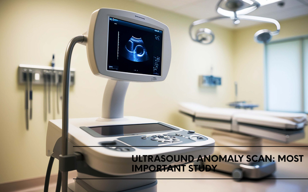
An ultrasound anomaly scan is a detailed medical examination that checks your baby’s development and screens for potential birth defects during pregnancy. This comprehensive guide is designed for expectant parents who want to understand this important prenatal test and make informed decisions about their care.
Getting an anomaly scan can feel overwhelming, especially when you’re not sure what to expect. We’ll walk you through the key aspects of this screening, including what conditions the scan can detect and when you should schedule your appointment for the best results. You’ll also learn what happens during the actual scan process and how to understand your results once they’re ready.
For parents in Mumbai seeking reliable prenatal care, centres like Sirona Shanti Diagnostics in Mahim and Prabhadevi offer advanced ultrasound services with experienced technicians who can guide you through this process with care and expertise.
Understanding What Ultrasound Anomaly Scans Detect
Major Structural Abnormalities in Organs and Limbs
Anomaly scans excel at detecting significant structural defects that affect the development of major organs and limbs. These comprehensive examinations can identify cleft lip and palate, limb deformities, including missing or shortened bones, and kidney abnormalities such as absent kidneys or cystic formations. The scan also reveals diaphragmatic hernias where abdominal organs move into the chest cavity, and gastroschisis or omphalocele where abdominal contents protrude outside the body wall.
Skeletal abnormalities become clearly visible during these detailed scans, including shortened limbs that might suggest dwarfism, spinal curvatures, and missing digits. Eye and facial malformations, along with cleft defects affecting the lip or palate, can be accurately identified when examining facial structures. These findings help medical teams prepare for potential surgical interventions or specialized care that might be needed after birth.
Heart Defects and Cardiovascular Issues
The four-chamber view of the heart represents one of the most critical aspects of anomaly scanning. Experienced sonographers can detect various congenital heart defects, including ventricular septal defects (holes between heart chambers), hypoplastic left heart syndrome, and transposition of the great arteries. The scan evaluates heart rhythm patterns, chamber sizes, and the proper positioning of major blood vessels.
Complex cardiac abnormalities such as tetralogy of Fallot, coarctation of the aorta, and valve stenosis can be identified through careful examination of blood flow patterns and structural anatomy. Early detection of these conditions allows families to connect with pediatric cardiologists and plan delivery at facilities equipped with specialised cardiac care units.
Neural Tube Defects Affecting Brain and Spine
Neural tube defects represent some of the most serious conditions detectable through anomaly scanning. Spina bifida, where the spinal column doesn’t close properly, can be identified along with its varying degrees of severity. Anencephaly, a condition where major portions of the brain and skull are missing, becomes clearly visible during scanning.
Encephalocele, where brain tissue protrudes through skull openings, and hydrocephalus, characterised by excess fluid in brain cavities, are additional neural tube conditions that skilled technicians at facilities like Sirona Shanti Diagnostics in Mahim can accurately identify. The scan also evaluates the cerebellum and measures various brain structures to ensure proper development. These findings help families understand the potential challenges ahead and connect with appropriate specialists.
Chromosomal Markers and Genetic Indicators
While anomaly scans cannot definitively diagnose chromosomal conditions, they can identify soft markers that suggest increased risk for genetic abnormalities. These markers include increased nuchal fold thickness, shortened femur length, bright spots in the heart or kidneys, and choroid plexus cysts in the brain.
The scan evaluates facial features for characteristics associated with Down syndrome, including a flattened facial profile and specific measurements. Growth restrictions, unusual fluid levels, and structural combinations can indicate various genetic syndromes. When multiple markers appear together, genetic counselling becomes valuable for understanding risks and exploring additional testing options. Expert sonographers in Prabhadevi and surrounding areas use advanced equipment to detect these subtle indicators accurately.
Optimal Timing for Maximum Detection Accuracy
Why 18-22 weeks offers the best window
The magic window for ultrasound anomaly scanning falls between 18 and 22 weeks of pregnancy for several crucial reasons. At this stage, your baby has developed enough for most major organs and structures to be clearly visible, yet still has sufficient space in the womb for optimal imaging. The baby’s bones haven’t hardened completely, allowing ultrasound waves to penetrate tissues more effectively and create clearer images.
During this timeframe, the amniotic fluid levels are ideal – not too little that would crowd the baby, and not excessive amounts that might create imaging challenges. This balance provides the perfect acoustic window for detailed examination of facial features, spine, heart chambers, kidneys, and limbs. Most importantly, if any anomalies are detected, there’s still adequate time for additional testing, consultations with specialists, and informed decision-making about the pregnancy’s future.
How baby size affects scan quality
Your baby’s size plays a critical role in determining scan quality and diagnostic accuracy. At 18-22 weeks, babies typically measure between 5.5 to 7 inches from crown to rump – large enough for detailed anatomical assessment but small enough to fit entirely within the ultrasound beam’s field of view.
Smaller babies (before 18 weeks) present challenges because their organs are still developing and may be too tiny to evaluate thoroughly. Conversely, larger babies (after 22 weeks) can create imaging difficulties as they begin to outgrow the optimal scanning window. Their increasing size means body parts may be positioned in ways that obscure other structures, and the hardening skeleton can cast shadows that interfere with the visualisation of internal organs.
The positioning also becomes more restrictive as pregnancy progresses. Babies have less room to move into ideal positions for scanning, making it harder to obtain the multiple views needed for comprehensive anomaly screening.
Benefits of scheduling during mid-pregnancy
Mid-pregnancy timing offers unique advantages that extend beyond just technical imaging considerations. Your energy levels are typically at their peak during the second trimester, making the appointment less physically demanding. Morning sickness has usually subsided, and the pregnancy fatigue of later stages hasn’t yet set in.
From a practical standpoint, scheduling during this window allows adequate time for follow-up procedures if needed. Should the scan reveal any concerns, there’s sufficient time for additional imaging, genetic counselling, specialist consultations, or amniocentesis if recommended. This timeline also provides families with the emotional space needed to process results and make informed decisions about their pregnancy journey.
For couples considering anomaly screening at reputable facilities in Mumbai’s Mahim or Prabhadevi areas, booking during this optimal window ensures access to the highest quality diagnostic imaging when it matters most. The combination of ideal fetal development, optimal imaging conditions, and adequate time for follow-up care makes the 18-22 week window the gold standard for anomaly detection.
What to Expect During Your Scan Appointment
Preparation steps for the clearest images
Your healthcare provider will ask you to drink several glasses of water about an hour before your appointment and avoid urinating until after the scan. This full bladder helps push your uterus upward and creates a clearer acoustic window for the ultrasound waves to travel through. The water acts like a natural lens, making your baby’s anatomy more visible and giving the sonographer better image quality.
Wear comfortable, loose-fitting clothes that can be easily lifted or removed from your abdomen. A two-piece outfit works best since you’ll only need to expose your belly area. Remove any jewellery around your waist or lower chest that might interfere with the examination. At facilities like Sirona Shanti Diagnostics in Mahim, staff will provide you with specific preparation instructions when you schedule your appointment.
Detailed examination process and duration
The anomaly scan typically takes 30 to 45 minutes, though complex cases or multiple babies may require longer. You’ll lie on an examination table while the sonographer applies warm gel to your abdomen. This gel eliminates air pockets between the ultrasound probe and your skin, ensuring clear sound wave transmission.
The sonographer will systematically examine your baby from head to toe, checking major organs and body systems. They’ll look at the brain structure, spine alignment, heart chambers and blood flow, kidneys and bladder, stomach and bowel, arms and legs including fingers and toes, and facial features. The examination follows standardized protocols to ensure nothing gets missed.
Real-time viewing of your baby’s development
Most facilities, including those in Prabhadevi area, position the monitor so you can watch the examination in real-time. You’ll see your baby moving, possibly sucking their thumb, or changing positions. The sonographer will point out different body parts and explain what they’re examining as they work through their checklist.
This becomes an exciting bonding experience for many parents. You might see your baby’s profile, watch their heartbeat on the screen, or catch them in an adorable position. Many parents describe feeling overwhelmed with emotion during these moments of connection with their unborn child.
Professional measurements and assessments
The sonographer takes precise measurements of your baby’s head circumference, abdominal circumference, thigh bone length, and other key structures. These measurements help determine if your baby is growing appropriately for their gestational age and can identify potential growth issues.
They’ll also assess amniotic fluid levels, placenta position, and umbilical cord insertion site. All findings are documented with still images and sometimes video clips. A radiologist or maternal-fetal medicine specialist reviews these images and measurements to provide your doctor with a comprehensive report about your baby’s development and well-being.
Interpreting Your Scan Results Effectively
Normal findings that indicate healthy development
When your ultrasound results show normal findings, you can feel confident that your baby is developing well. A healthy anomaly scan typically reveals proper organ formation, with all four chambers of the heart visible and functioning normally. The spine should appear straight and complete, while the brain shows typical development with visible cerebral hemispheres and proper skull formation.
Your baby’s limbs should be present and proportionate, with clear visualization of arms, legs, hands, and feet. The kidneys should be visible on both sides, and the stomach should appear as a fluid-filled structure. Normal growth measurements include head circumference, abdominal circumference, and femur length that fall within expected ranges for gestational age.
Other reassuring signs include appropriate amniotic fluid levels (neither too much nor too little), a properly positioned placenta, and normal umbilical cord insertion. The baby’s movements during the scan, while sometimes making imaging challenging, actually demonstrate healthy neurological development.
Soft markers versus significant abnormalities
Understanding the difference between soft markers and significant abnormalities helps you process your scan results appropriately. Soft markers are minor variations that appear on ultrasound but often resolve on their own or have minimal clinical significance. These might include echogenic intracardiac focus (bright spots in the heart), choroid plexus cysts (small fluid-filled spaces in the brain), or slightly shortened nasal bone measurements.
Soft markers don’t typically require immediate intervention and are found in many healthy pregnancies. However, they may slightly increase the statistical risk for certain conditions, which is why your healthcare provider discusses them with you.
Significant abnormalities represent structural defects or major developmental issues that require medical attention. These include heart defects, neural tube defects like spina bifida, kidney abnormalities, or limb malformations. Unlike soft markers, these findings usually need follow-up care, additional testing, or treatment planning.
Your doctor at facilities like Sirona Shanti Diagnostics in Mahim or Prabhadevi will explain which category your findings fall into and what this means for your pregnancy moving forward.
When additional testing may be recommended
Additional testing becomes necessary when scan results raise questions that need clearer answers. If structural abnormalities are detected, you might need specialized scans performed by maternal-fetal medicine specialists who have advanced training in high-risk pregnancies.
Detailed cardiac scans may be recommended if heart abnormalities are suspected. These specialized ultrasounds provide more comprehensive views of your baby’s heart structure and function. Similarly, if brain abnormalities are noted, fetal MRI might offer better soft tissue detail than ultrasound alone.
Genetic testing options include amniocentesis or chorionic villus sampling (CVS) when chromosome abnormalities are suspected. Your doctor might also suggest cell-free DNA testing from your blood, which screens for common genetic conditions with high accuracy.
Follow-up scans are often scheduled to monitor development over time. Some conditions that appear concerning early in pregnancy may improve or resolve as the baby continues growing. Regular monitoring helps your healthcare team track these changes and adjust care plans accordingly.
Your medical team will discuss all testing options, explaining the benefits, risks, and limitations of each procedure to help you make informed decisions about your pregnancy care.
Managing Emotions and Next Steps After Results
Processing unexpected findings with support
Receiving unexpected results from your anomaly scan can feel overwhelming and isolating. The range of emotions you might experience is completely normal – shock, fear, sadness, anger, or confusion are all valid responses. Many parents describe feeling like time stopped when they first heard concerning news about their pregnancy.
Your healthcare team understands these feelings and should provide immediate emotional support alongside medical information. Don’t hesitate to ask for clarification if medical terms seem confusing or if you need information repeated. Having a support person with you during these conversations can help you process information better and remember important details later.
Professional counselling services are often available through your healthcare provider or can be arranged quickly. These specialists help couples navigate the complex emotions and decisions that may lie ahead. Support groups, either in-person or online, connect you with other parents who have faced similar situations and can offer valuable perspectives.
Remember that taking time to process information is important. You don’t need to make immediate decisions about your pregnancy in most cases. Quality healthcare facilities like those in Mahim and Prabhadevi often have dedicated counselling teams who specialise in supporting families through these challenging moments.
Follow-up care options and specialist referrals
When your anomaly scan reveals potential concerns, your care team will typically recommend additional testing or specialist consultations. The type of follow-up depends on the specific findings and may include more detailed ultrasounds, genetic testing, or consultations with pediatric specialists.
Maternal-fetal medicine specialists provide expert evaluation of complex cases and can offer more detailed imaging using advanced ultrasound equipment. These specialists work closely with your regular obstetrician to create comprehensive care plans tailored to your specific situation.
Some conditions may require consultations with pediatric cardiologists, neurologists, or surgeons who can explain potential treatments available after birth. These meetings help you understand what to expect and allow you to prepare mentally and practically for your baby’s arrival.
Additional diagnostic tests might include amniocentesis or more detailed ultrasounds at specialized centers. Reputable diagnostic centers in areas like Prabhadevi often have state-of-the-art equipment and experienced sonographers who can provide clearer pictures of your baby’s development.
Making informed decisions about your pregnancy
Armed with complete information from your medical team, you’ll need to consider various options for moving forward with your pregnancy. This decision-making process is deeply personal and should never feel rushed or pressured by anyone else’s opinions or timelines.
Understanding the full spectrum of your baby’s condition helps you make choices aligned with your family’s values and circumstances. Some conditions detected during anomaly scans are minor and easily treatable after birth, while others may require ongoing medical care or have more serious implications.
Consider practical factors alongside emotional ones – your support system, financial resources, existing children, and personal beliefs all play important roles in your decision-making process. Speaking with families who have children with similar conditions can provide valuable real-world perspectives on daily life and care requirements.
Your medical team should present all available options clearly, including continued pregnancy with specialized care planning, additional monitoring, or pregnancy termination if appropriate and desired. Take advantage of resources available through established healthcare center like Sirona Shanti Diagnostics which often provide comprehensive counseling services to help families navigate these difficult decisions with confidence and support.
The anomaly scan stands as one of pregnancy’s most important milestones, offering expecting parents valuable insights into their baby’s development around 18-20 weeks. This detailed ultrasound checks for structural abnormalities, measures growth, and provides reassurance during a time filled with anticipation. Understanding what the scan can and cannot detect helps you prepare mentally and sets realistic expectations for this significant appointment.
Remember that most scans reveal healthy, developing babies, and even when concerns arise, early detection opens doors to better planning and care options. Trust your healthcare team to guide you through the process, ask questions when you need clarity, and lean on your support network during this emotional journey. Whether your results bring relief or require additional follow-up, you’re taking an important step in ensuring the best possible outcome for your growing family.
Dr. Kunaal Jain
Consultant Radiologist
───────
Go to Our Website and Get in Touch
Don’t ignore what your body is trying to tell you. A simple thyroid test can help you feel like yourself again, whether you’ve gained or lost weight for no reason or are always tired. To find out more about our full range of health services and packages.
Go to our website at https://sironashanti.com
OR
Call +91 98191 88333 or WhatsApp +91 86554 08047.
───────
Blog Keywords:
Hysterosalpingogram, HSG test, fallopian tube blockage, fertility evaluation, uterine abnormalities, HSG procedure, Sironashanti Diagnostics, fertility test, tubal patency test, blocked fallopian tubes, HSG contrast dye, uterine fibroids HSG, hydrosalpinx diagnosis, Asherman’s syndrome, HSG pain management, HSG recovery, female infertility testing. Ultrasound anomaly scan, pregnancy ultrasound, birth defect screening, prenatal test, baby development scan, Sironashanti Diagnostics, Mahim anomaly scan, Prabhadevi ultrasound services, Mumbai prenatal care, advanced fetal imaging, anomaly scan procedure, fetal anomaly screening.
Common Keywords:
Best NABL accredited pathology lab in Mumbai, Best pathology lab in Mahim Dadar Prabhadevi Worli, Best radiology centre center in mumbai, Best sonography center in mumbai, Best Radiologist / Sonologist in Mahim Worli Dadar Prabhadevi, Best Cardiologist in Mumbai, best cardiology centre, centre in Mahim Dadar Prabhadevi, ecg at home, xray at home, blood collection at home, blood collection in mumbai, bloold collection south mumbai, verified best diagnostics centers, sironashanti diagnostics center
Common Extra Keywords:
diagnostic centre Mumbai, best diagnostic centre Mumbai, pathology lab Mumbai, best pathology lab Mumbai, NABL accredited lab Mumbai, diagnostic centre near me Mumbai, trusted diagnostic centre Mumbai, Sirona Shanti Diagnostics, affordable diagnostic centre Mumbai, diagnostic centre open Sunday Mumbai, diagnostic centre Prabhadevi, pathology lab Prabhadevi, blood test Prabhadevi Mumbai, diagnostic centre Mahim, pathology lab Mahim, blood test Mahim Mumbai, diagnostic centre Dadar, pathology lab Dadar West, blood test Dadar Mumbai, diagnostic centre Bandra, pathology lab Bandra West, blood test Bandra Mumbai, diagnostic centre Lower Parel, health checkup Lower Parel Mumbai, diagnostic centre Worli, blood test Worli Mumbai, home blood collection Mumbai, home blood test Colaba, home sample collection South Mumbai, home blood collection Andheri, home blood test Juhu, home blood collection Santacruz, home blood test Vile Parle, home blood collection Khar Mumbai, at home blood test Mumbai, blood sample collection at home Mumbai, full body checkup Mumbai, full body health package Mumbai, full body blood test Mumbai, complete blood count Mumbai, CBC test Mumbai, thyroid test Mumbai, thyroid profile Mumbai, TSH test Mumbai, diabetes test Mumbai, HbA1c test Mumbai, blood sugar test Mumbai, lipid profile test Mumbai, cholesterol test Mumbai, liver function test Mumbai, kidney function test Mumbai, urine test Mumbai, Vitamin D test Mumbai, Vitamin B12 test Mumbai, dengue test Mumbai, malaria test Mumbai, typhoid test Mumbai, ECG test Mumbai, ECG near me Mumbai, 2D echo test Mumbai, echocardiogram Mumbai, stress test Mumbai, TMT test Mumbai, heart checkup Mumbai, cardiac test Mumbai, digital X-ray Mumbai, X-ray centre Mumbai, X-ray near me Mumbai, sonography Mumbai, ultrasound test Mumbai, USG scan Mumbai, mammography Mumbai, breast screening Mumbai, home ECG Mumbai, home X-ray Mumbai, blood test near me Mumbai, lab test near me Mumbai, pathology lab near me, diagnostic centre near me, full body checkup near me Mumbai, health checkup near me Mumbai, best lab near me Mumbai, blood collection near me Mumbai, ECG near me, sonography near me Mumbai, health checkup packages Mumbai, annual health checkup Mumbai, preventive health checkup Mumbai, women health checkup Mumbai, senior citizen health checkup Mumbai, PCOD test Mumbai, hormonal test Mumbai, fertility test Mumbai, thyroid specialist Mumbai, endocrinology clinic Mumbai, gynaecology consultation Mumbai, cardiology consultation Mumbai, how much does full body checkup cost in Mumbai, best full body checkup package Mumbai affordable, same day blood test report Mumbai, online report delivery diagnostic Mumbai, blood test report on WhatsApp Mumbai, NABL lab near me Mumbai, ISO certified pathology lab Mumbai, accurate blood test Mumbai, trusted blood test results Mumbai, walk-in diagnostic centre Mumbai.